
Magnetic Resonance Imaging (MRI) is a medical imagery technique that does not expose the body to ionizing radiation (such as X-ray). Instead, it relies on a strong magnetic field to excite atoms in the tissues, more specifically hydrogen atoms present in water (water accounts for 70% of the human body mass). Measuring the rate at which the atoms go back to their equilibrium state allows to reconstruct the spatial distribution of water, and by extension to differentiate tissue types.Finding the magnetic field that maximizes the contrast between two types of tissue can be written as an optimal control problem, studied for instance in [1,2]. The magnetization vector 

with u the magnetic field (control) and 

Considering two different particles with spins 


with the final time set to a multiple of the minimum time 

We illustrate this problem on the (cerebro-spinal fluid,water) case, see below, with an example of a 2BS structure.
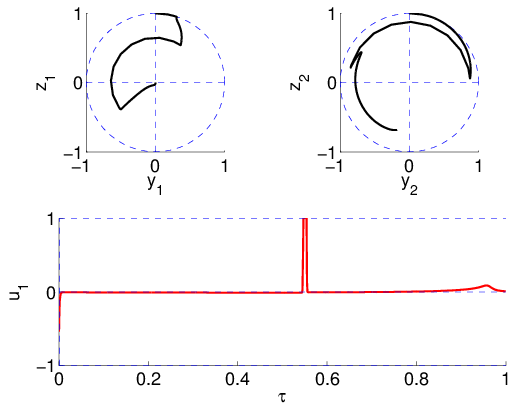
MRI: contrast by saturation, (cerebro-spinal fluid,water) case.
A comprehensive study using indirect shooting and differential continuation methods (HAMPATH,[3]) indicates the existence of numerous families of local solutions with different structures, and shows that the optimal structure depends on the final time, as shown here.
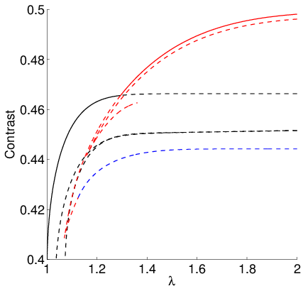
Contrast: branches of local solutions (1BS: black, 2BS: blue, 3BS: red)
References
[3] O. Cots. Controle optimal geometrique: methodes homotopiques et applications. PhD thesis, 2012.